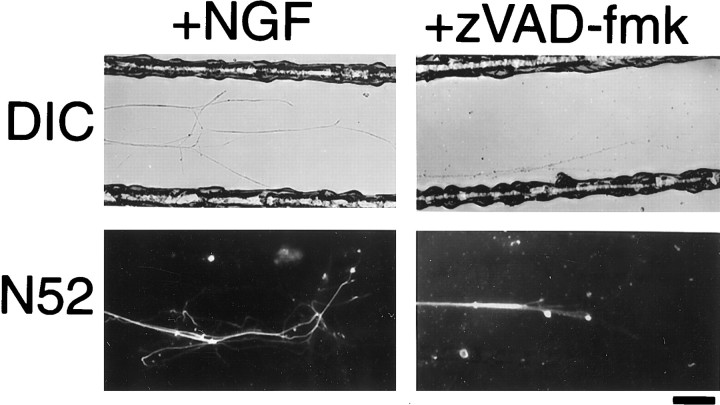
Fig. 7.
DIC and immunofluorescence micrographs showing the effect of caspase inhibitors on axons locally deprived of NGF. P0 DRG neurons were dissociated and plated in the central compartment of a three-chambered culture dish, with NGF in all of the compartments. After 7 d, the neurons had extended axons into the two distal compartments, and the medium in some of the right-hand distal chamber was replaced with medium that lacked NGF and contained both anti-NGF antibodies and zVAD-fmk. Four days later, the neurons were fixed and then studied by DIC microscopy or stained with the N52 anti-neurofilament antibody and then studied with confocal immunofluorescence microscopy. Many of the axons in compartments deprived of NGF and treated with zVAD-fmk (shown) degenerated; axons in compartments deprived of NGF without zVAD-fmk (data not shown) showed comparable degeneration. Different fields are shown for DIC and immunofluorescence images. Scale bar, 100 μm.

