
Fig. 3.
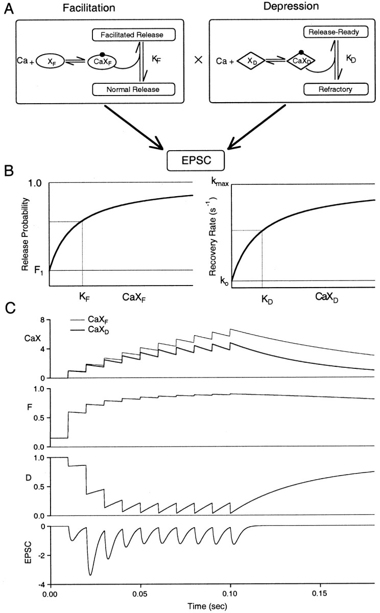
FD model for Ca-dependence of short-term plasticity. A, Left, Residual presynaptic calcium binds to site XF, and the complex CaXF then binds to the release site causing an enhancement of release probability. Right, Schematic of residual presynaptic calcium binding to site XD, which then binds with the refractory release site driving a transition back to the release-ready state. B, Left, F plotted as a function of CaXF ranging from a minimal probability of F1 (no residual calcium) to a maximum of 1. The dissociation constant for CaXF is KF.Right, the recovery rate for depression is plotted as a function of CaXD with a minimum rate of ko, a maximum rate of kmax, and CaXDdissociation constant KD. C, Presynaptic levels of CaXF (thin line) and CaXD (thick line), fraction of available synapses that undergo release (F), fraction of release-ready synapses (D), and normalized EPSC during a train of 10 stimuli at 100 Hz. Model parameters for this simulation were ρ = 3.4, F1 = 0.15, τF = 100 msec, τD = 50 msec, kmax = 30 sec−1, ko = 2 sec−1, KD = 2. Note that CaXF and CaXD were normalized to their respective dissociation constants.