
Fig. 1.
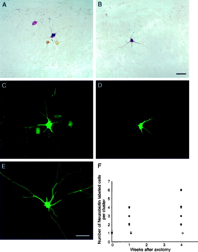
Axotomized motor neurons are extensively dye-coupled. A, Single plane projection of confocal stack of images showing a cluster of Neurobiotin-labeled motor neurons after injection of a single medial gastrocnemius motor neuron in a cat axotomized 1 week previously. Neurobiotin was revealed with HRP-conjugated streptavidin and a chromogenic reaction for HRP. There were a total of four labeled cells in this cluster, which occupied a region 214 × 234 × 189 μm in the dorsoventral, mediolateral, and rostrocaudal dimensions, respectively.B, Single plane projection of confocal stack of images showing a single cell body and a portion of the dendritic arbor of an injected peroneous motor neuron from a cat axotomized 1 week previously. Neurobiotin was revealed with fluorescein-conjugated streptavidin (also C–E). The absence of dye coupling in nonaxotomized motor neurons in animals in which the gastrocnemius–soleus nerve had been severed argues that gap junctional coupling is only observed among injured neurons. Scale bar:A, B, 100 μm. C, Single plane projection of confocal stack of images showing a cluster of Neurobiotin-labeled motor neurons after injection of a single medial gastrocnemius motor neuron in a cat axotomized 4 weeks previously. There were a total of four labeled cells in this cluster, which occupied a region 260 × 220 × 125 μm in the dorsoventral, mediolateral, and rostrocaudal dimensions, respectively.D, Single plane projection of confocal stack of images showing a single cell body and a portion of the dendritic arbor of an injected, nonaxotomized peroneous motor neuron from a cat axotomized 4 weeks previously. The absence of dye coupling in nonaxotomized motor neurons in animals in which the gastrocnemius–soleus nerve had been severed argues that gap junctional coupling is only observed among injured neurons, even at long times after axotomy. Scale bar:C–E, 100 μm. E, Single plane projection of confocal stack of images showing a single cell body and a portion of the dendritic arbor of an injected medial gastrocnemius motor neuron from a normal adult cat, showing the absence of dye coupling. F, Summary of the number of Neurobiotin-labeled cells per cluster in normal spinal cord in spinal cord 1 and 4–6 weeks after axotomy (filled circles) and in nonaxotomized motor pools in spinal cord 1 and 4–6 weeks after axotomy (open circles).