
Fig. 5.
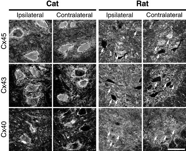
Connexin protein expression in motor neurons is unchanged after axotomy in rat and cat spinal cord. To determine whether connexin proteins are expressed in rat and cat motor neurons, immunostaining with antibodies specific for Cx45, Cx43, and Cx40 was performed in cat (axotomized side, left; contralateral side, left middle) and rat (axotomized side, right middle; contralateral side,right) spinal cord. Shown are single plane projections of confocal stacks of images from cat L6–L7 dorsolateral ventral spinal cord containing the gastrocnemius motor pools or rat L3–L6 lateral and ventral spinal cord containing the sciatic nerve motor pools, obtained on a Leica TCS-4D system with a 40×, 1.25 NA oil immersion lens. In both rat and cat, anti-Cx45 (top row), Cx43 (middle row), and Cx45 (bottom row) antibodies revealed punctate staining surrounding motor neuron soma (several are indicated with arrows in each rat panel) and primary dendrites in the ventral horn. Some motor neurons had prominent cytoplasmic staining in addition to punctate, membrane-associated staining. Similar patterns of staining were observed in unmanipulated control animals (data not shown) and were similar in the spinal cord ipsilateral and contralateral to sciatic nerve cut 1 and 4–6 weeks after axotomy. In the middle row, note the absence of a change in Cx43 protein expression in glia in and around axotomized motor neurons (but see Rohlmann et al., 1993, 1994). These results show that, in rat and in cat, axotomy does not result in a detectable change in connexin protein expression in injured motor neurons. Scale bar, 100 μm.