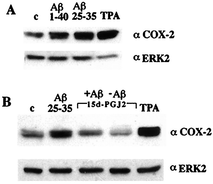
Fig. 7.
Aβ-stimulated COX-2 expression in THP-1 cells is inhibited by PPARγ agonists. A, THP-1 monocytes were incubated alone (c), with fibrillar Aβ25–35 (60 μm) or Aβ1–40 (60 μm) in suspension, or with 100 nm TPA for 18 hr in serum-free RPMI, and COX-2 expression was monitored by Western analysis of cellular lysates using an anti-COX-2-specific antibody. The blots were reprobed using an anti-ERK2 antibody as a control for protein loading. B,THP-1 cells were incubated in vehicle alone (c; DMSO) or stimulated with fibrillar Aβ25–35 (60 μm) or TPA (100 nm) in the presence or absence of the PPARγ agonist 15d-PGJ2 (50 μm).

