
Fig. 2.
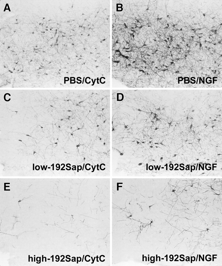
Loss of p75NGFr-immunoreactive CBF neurons after immunotoxic lesioning accompanied by NGF-induced hypertrophic changes. Representative photomicrographs of the anterior part of the NBM show the different degrees of diminished p75NGFr-immunoreactive neurons after injection of PBS (A), 1 μg of 192Sap (C), and 2.7 μg 192Sap (E). The area of p75NGFr-immunoreactive neurons as well as the density of neuritic sprouting are increased in the PBS/NGF animals (B) but also in the immunotoxic lesioned animals independent of the remaining number of p75NGFr-immunoreactive neurons (D, F).