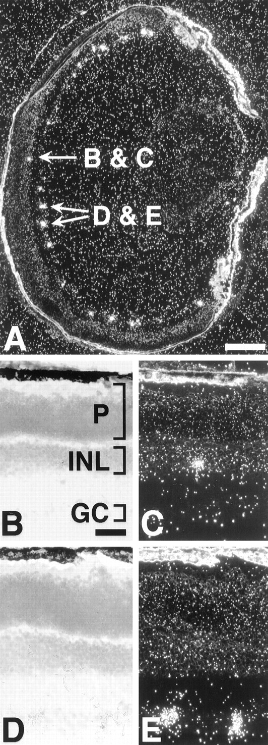
Fig. 5.

Melanopsin is expressed in the mouse inner retina.A, Cross-section of a 10-d-old mouse eye probed with an antisense mouse melanopsin riboprobe. B,C, Bright-field and dark-field photomicrographs of indicated cell within the amacrine cell layer in A.D, E, Bright-field and dark-field photomicrographs of indicated cell pair within the ganglion cell layer in A. GC, Ganglion cell layer;INL, inner nuclear layer; P, photoreceptor layer. Scale bars: A, 250 μm;B, 50 μm.
