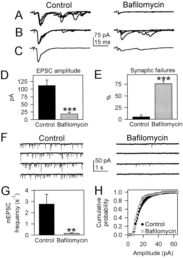
Fig. 1.
Bafilomycin A1 reduced the probability of synaptic transmitter release. A,B, Representative traces of evoked EPSCs in control cultures and after incubation with bafilomycin. In many cases, EPSCs were absent in bafilomycin-treated cultures. When present, the amplitude of EPSCs in bafilomycin was smaller than in control, and the number of synaptic failures was increased by bafilomycin. C, Averaged (n = 10) EPSCs in control and bafilomycin. Shown is the same pair of pre-postsynaptic neurons as in B. D,Mean evoked EPSC amplitude in control and bafilomycin-treated cultures (n = 25 and 35 presynaptic neurons, respectively).E, Mean percentage of synaptic failures observed during trials of 10 stimuli delivered at 1 Hz in control and after incubation with bafilomycin (n = 25 and 35 presynaptic neurons, respectively). F, mEPSCs in control and after incubation with bafilomycin. G, Mean frequency of mEPSCs measured during 1–4 min in control and in bafilomycin-treated cells (n = 9). H, Average cumulative probability plots of the mEPSC amplitude in control (n = 9) and after incubation with bafilomycin (n = 8). Cumulative probability plots were calculated in 1 pA bins and were not significantly different between control and bafilomycin-treated cells (Kolmogorov–Smirnov test). Holding potential was −60 mV. Significant differences with respect to control were established by the Student's t test atp < 0.02 (**) and p < 0.001 (***).

