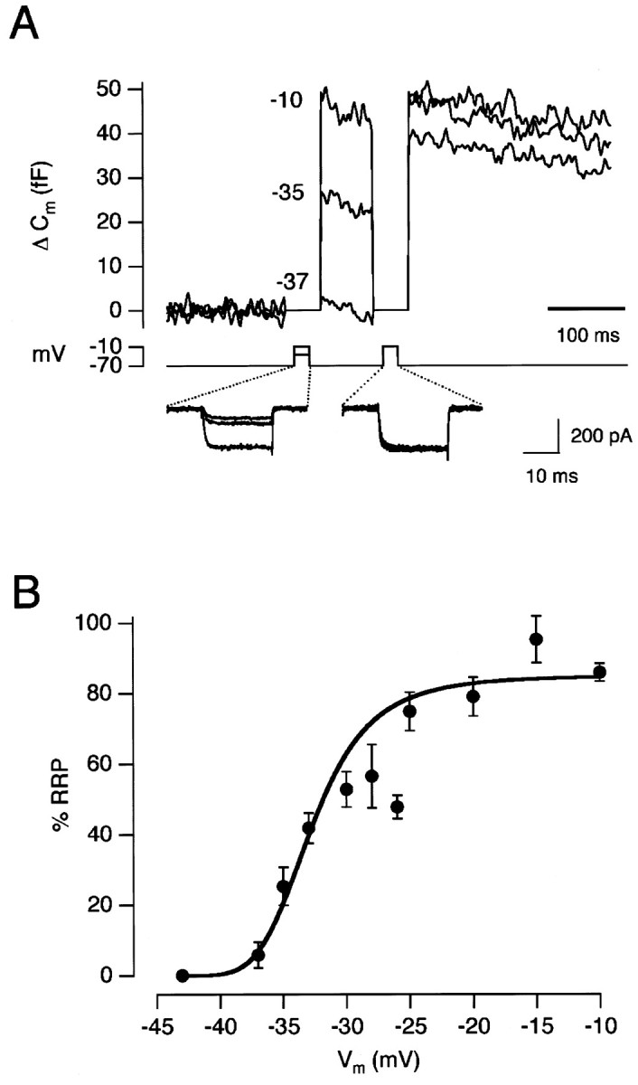
Fig. 8.

Voltage-dependence of exocytosis for 20 msec pulses. A, Capacitance increases elicited by 20 msec depolarizations to −37, −35, and −10 mV. Each test stimulus was followed by the emptying stimulus. The Ca2+ currents for each stimulus are shown below. B, The amount of exocytosis elicited by a 20 msec depolarization to various membrane potentials. Each point is the average of 4–16 measurements. Results collected from a total of 20 cells. The capacitance increase to the test stimulus was expressed as a percentage of the RRP. The line fitted through thepoints is a Hill function (see Results).
