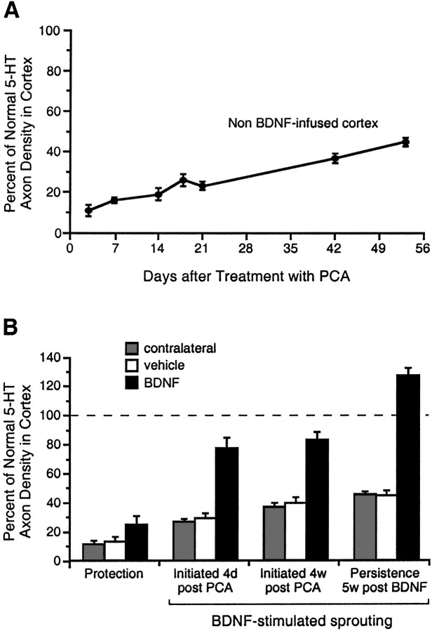
Fig. 3.
The density of serotonin axons (SERT-immunoreactive) in frontoparietal cortex of PCA-lesioned animals and expressed as a percentage of the normal density found in the homologous cortex of intact animals. A, The normally slow reinnervation of cortex by 5-HT axons after PCA administration, as determined in the contralateral cortex of vehicle- and BDNF-infused animals (the two measures were not significantly different at any time point, and were thus collapsed for presentation). B,Effects of vehicle or BDNF infusions (12 μg/d) on PCA-lesioned 5-HT axons (measured locally at the infusion site), as investigated in the following treatment paradigms: Protection (protocol, Fig.1C; leftmost set of histograms); Sprouting (protocol, Fig. 1D) where 2 week BDNF infusions were initiated at 4 d or 4 weeks after PCA administration (center two sets of histograms, respectively); and Persistence of sprouted axons (protocol, Fig. 1E) at 5 weeks after terminating the BDNF infusion (rightmost set of histograms). Gray histogram bars, The contralateral cortex of vehicle- and BDNF-infused animals (the two values were not significantly different in any treatment paradigm, and were thus pooled); white bars,vehicle infusion in the ipsilateral (right) cortex; black bars, BDNF infusion. In all treatment paradigms, the 5-HT axon density was higher in the BDNF-infused cortex relative to the vehicle-infused and contralateral cortex (ANOVA followed by the Newman–Keuls multiple range test, p < 0.05), whereas the control conditions did not differ significantly (p > 0.05).

