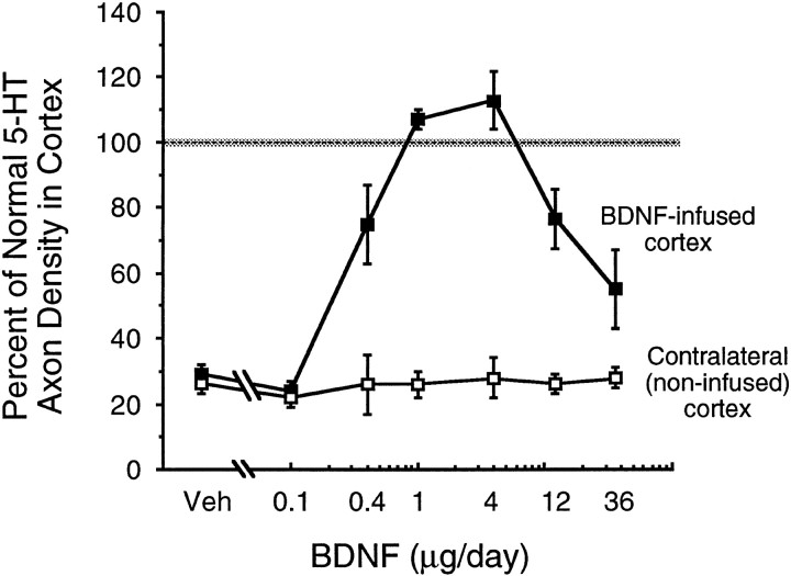
Fig. 5.
The density of serotonin axons (SERT-immunoreactive) after intracortical infusion of vehicle or 0.1, 0.4, 1, 4, 12, or 36 μg/d of BDNF (protocol, Fig.1D, 2 week infusions were started 4 d after PCA); measured in the area of BDNF-positive immunoreactivity (determined in adjacent sections) and expressed as a percentage of the normal density in intact animals. Closed squares,Vehicle or BDNF infusions in the ipsilateral (right) cortex;open squares, the contralateral (noninfused) side of cortex. ANOVA followed by the Bonferonni post hoc test revealed that 5-HT axon density was significantly higher after 0.4–12 μg/d of BDNF relative to vehicle; the 12 and 36 μg/d doses of BDNF resulted in a lower density than the 4 μg/d dose (F(6,24) = 15; p < 0.0001).

