
Fig. 6.
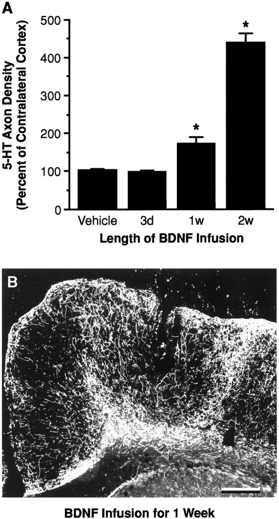
The serotonergic innervation after infusion of vehicle or BDNF (4 μg/d) for 3, 7, or 14 d in cortex of previously PCA-lesioned animals. A, SERT axon density was measured at the BDNF infusion site and expressed as a percentage of the density in the contralateral cortex (noninfused but PCA-lesioned). Treatment paradigms are described in Materials and Methods (Experimental paradigm). *p < 0.05, relative to the vehicle-infused and contralateral cortex (ANOVA followed by the Newman–Keuls multiple range test). B, Dark-field photomicrograph of 5-HT-immunoreactive axons after a 1 week BDNF infusion (4 μg/d; initiated at 7 d after PCA administration) in cortex. The effects of vehicle and 2 week BDNF (4 μg/d) infusions on 5-HT axons (using a similar treatment protocol) are shown in Figures2F and 4E, respectively. Scale bar, 0.5 mm.