
Fig. 7.
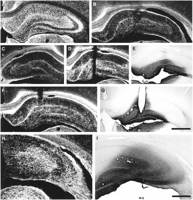
The serotonergic innervation in dorsal hippocampus after local infusion of vehicle or BDNF (4 μg/d) in PCA-lesioned animals (protocol, Fig. 1D, 2 week infusions were started 1 week after PCA). A–D, F, H,5-HT-immunoreactive axons in hippocampus (dark-field photomicrographs; coronal sections); E, G, I, area of BDNF diffusion as determined by BDNF immunocytochemistry in adjacent sections (bright-field photomicrographs). A, The normal 5-HT innervation in hippocampus of intact rats. B, Vehicle infusion in the PCA-lesioned hippocampus (note cannula tract at center). C, The extent of 5-HT denervation normally seen 3 weeks after PCA administration in the contralateral hippocampus.D, E, Sprouting of 5-HT axons in the dentate gyrus (D) after local infusion of BDNF (E). F, G, Sprouting of 5-HT axons in CA1 and the dentate gyrus (F) in response to BDNF infusion (G). H, I, Sprouting of 5-HT axons in CA3 (H) after infusion of BDNF (I); note the higher magnification inH and I than in A–G. Scale bars: A–G (shown in G), 1 mm;H, I (shown in I), 0.5 mm.