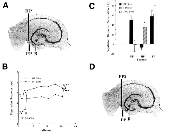
Fig. 3.
Lack of reciprocal potentiation of hilar and perforant path granule cell inputs. To test the hypothesis that hilar path (HP) and perforant path (PP) afferents independently excite granule cells, LTP of each pathway (n = 6) was induced with a tetanus, and changes were evaluated in the other pathway. Asterisk signifies statistical significance at p < 0.05 level.A, Scanned cresyl violet-stained guinea pig hippocampal slice with a schematic of the recording electrode (R) in the granule cell layer (G), and hilar path (HP) and perforant path (PP) stimulating electrodes in the molecular layer (M) of the dentate gyrus.B, Representative example of granule cell population responses to stimulation of hilar (HP Stim) and perforant (PP Stim) pathways before and 30 min after tetanic stimulation of the hilar path (HP Tetanus). In contrast to the sustained potentiation seen in the tetanized HP pathway (HP Stim), no change was evident in the perforant path (PP Stim). C, Tetanization of the perforant pathway (PP Tetanus) produced significantly more potentiation at 30 min (LTP) in the perforant path (PP Stim) than in the hilar path (HP Stim). Conversely, tetanization of the hilar path (HP Tetanus) produced significantly more LTP in the hilar path (HP Stim) than in the perforant path (PP Stim). In contrast to these results, responses to perforant path stimulation (PP Stim) after LTP induction (PP Tetanus) did not significantly differ from responses to a second stimulating electrode (PP2 Stim) placed in the perforant path (as in D). D, Scanned cresyl violet-stained guinea pig hippocampal slice schematic of the recording electrode (R) in the granule cell layer (G) and two perforant path (PP,PP2) stimulating electrodes in the molecular layer (M) of the dentate gyrus.

