
Fig. 1.
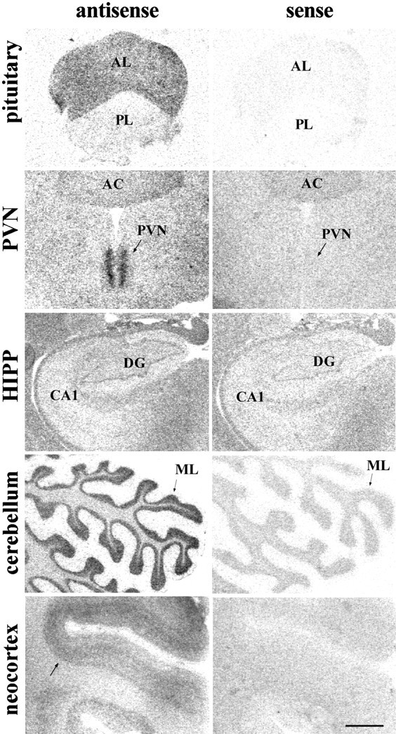
Left, Representative autoradiograms showing GR mRNA in situ hybridization signal with the [35S]-labeled human cRNA antisense probe in the rhesus macaque pituitary and several regions of the CNS. Note the low hybridization levels obtained in DG and CA1–4 of the hippocampal formation, in comparison to the other regions sampled.Right, Hybridization signal detected when using the sense cRNA radiolabeled probe in adjacent sections. Scale bar, 1.5 mm.AC, Anterior commissure; AL, anterior lobe of the pituitary; BNST, bed nucleus of the stria terminalis; CA1–4, Cornu Ammonis subfields (1–4);CBL, cerebellum; CEA, central nucleus of amygdala; DG, dentate gyrus of the hippocampus;Ent ctx, Entorhinal cortex; GL, cerebellar cortex, granular layer; HIPP, hippocampal formation; ML, cerebellar cortex, molecular layer;MEA, medial nucleus of amygdala; P, cerebellar cortex, Purkinje cell layer; PIT, pituitary;PL, posterior lobe of the pituitary;PreS, presubiculum; ProS, prosubiculum;PVN, hypothalamic paraventricular nucleus;SON, supraoptic nucleus; Sub, subiculum;VMH, ventromedial hypothalamic nucleus;V, third ventricle.