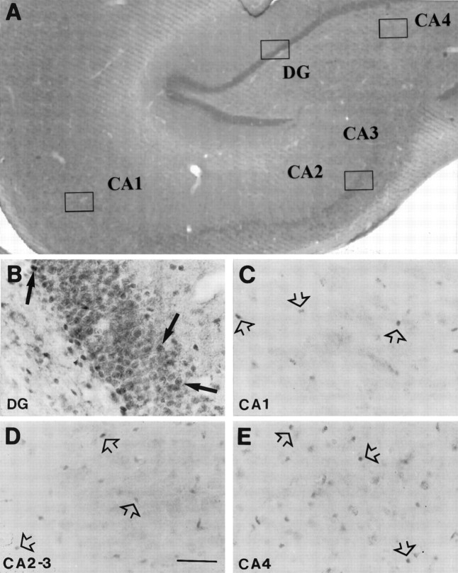
Fig. 4.
Photomicrographs of coronal sections through the macaque hippocampal formation, where a very low density of moderately GR-IR cells was observed. A, Low magnification view of the primate hippocampus; B, granule cells in the DG, where some moderately stained GR-IR nuclei were found (arrows); representative images of CA1 (C), CA2–3 (D), and CA4 (E) subfields, showing a very low density of very lightly stained GR-immunoreactive nuclei whose predominant small nuclear profile size could belong to glial cells (arrowheads). Scale bar (applies toB–E), 50 μm; 40×.

