
Fig. 9.
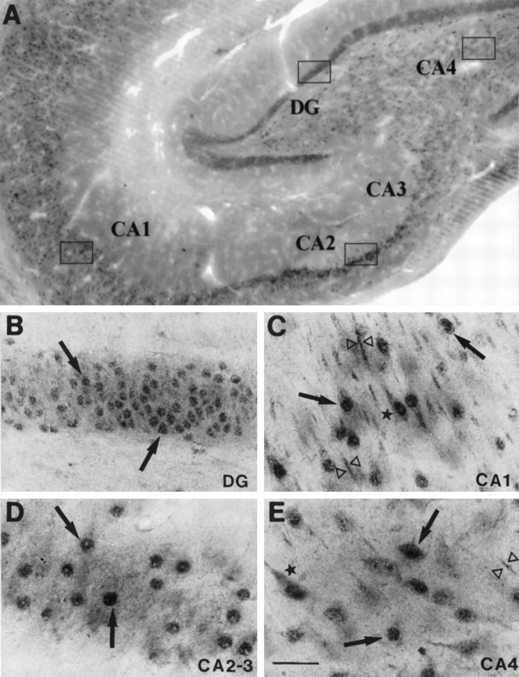
Immunohistochemical localization of MR in the rhesus monkey hippocampal formation. A, Low magnification field of view; B, high density of moderately MR-IR nuclei (arrows) within the DG granule cell layer; C, high density of MR-IR cells, moderately immunostained, in the CA1 subfield, where weak cytoplasmic (star) and dendritic (open triangles) immunostaining was detected in addition to the predominant nuclear immunoreactivity (arrows); E, this same pattern was observed in the CA4 pyramidal cell layer, although less density of MR-IR cells was detected; D, intensely MR-IR cells in the pyramidal cell layer of CA2 and CA3. Scale bar, 50 μm.