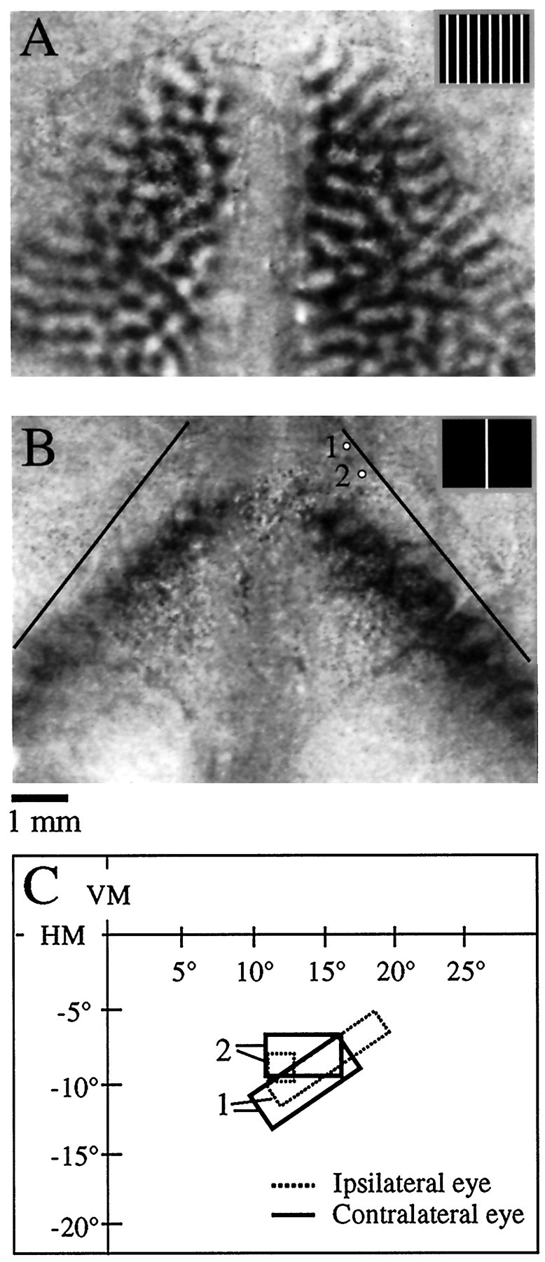
Fig. 2.

Bilateral orientation difference signal and bilateral response to a stimulus at the VM (animal TS9751).A, Bilateral orientation difference signal. Dark areas of the image were preferentially activated by a vertical grating (stimulus shown in inset), and white areas were preferentially activated by a horizontal grating.B, Bilateral pattern of activity in response to a bar stimulus (0.5° wide moved in a 2° wide window placed at the VM, shown in inset). Dark areas of the image were strongly activated by the bar stimulus as compared to a blank screen. Thethin black lines denote the V1/V2 border as defined by the orientation signal shown in A. Note that the cortical representation of the VM is displaced from the V1/V2 border in each hemisphere, implying the existence of a representation of the ipsilateral visual field. Scale bar applies to A andB. C, Multiunit receptive field plots for the two recording sites depicted in B. The ipsilateral (dashed lines) and contralateral eye (thick lines) receptive fields are overlapping at each location, indicating minimal misalignment of the eyes. The receptive fields are located in the right (ipsilateral) visual field, consistent with the optical imaging results.
