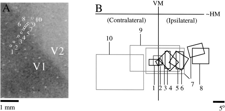
Fig. 5.
Physiological confirmation of the ipsilateral visual field representation (animal TS9756). A, Location of 10 recording sites relative to a Nissl-stained section cut tangentially through layer 4; the darkly stained region of the tissue corresponds to V1. B, Multiunit receptive field plots for the recording locations shown in A. For site 1 the contralateral (thick black line) and ipsilateral eye (dashed line) receptive fields were determined separately to confirm proper eye alignment. Receptive fields for all other sites were plotted using the contralateral eye only. Receptive fields for sites that appeared to be in V1 are shown inblack, and receptive fields for sites that appeared to be in V2 are shown in gray. At site 7, recordings were made at two different depths, and receptive fields from both depths are shown. Based on these recordings, the V1/V2 border appeared to be near sites 7 and 8, which corresponds to the position of the border on the Nissl-stained section.

