
Fig. 6.
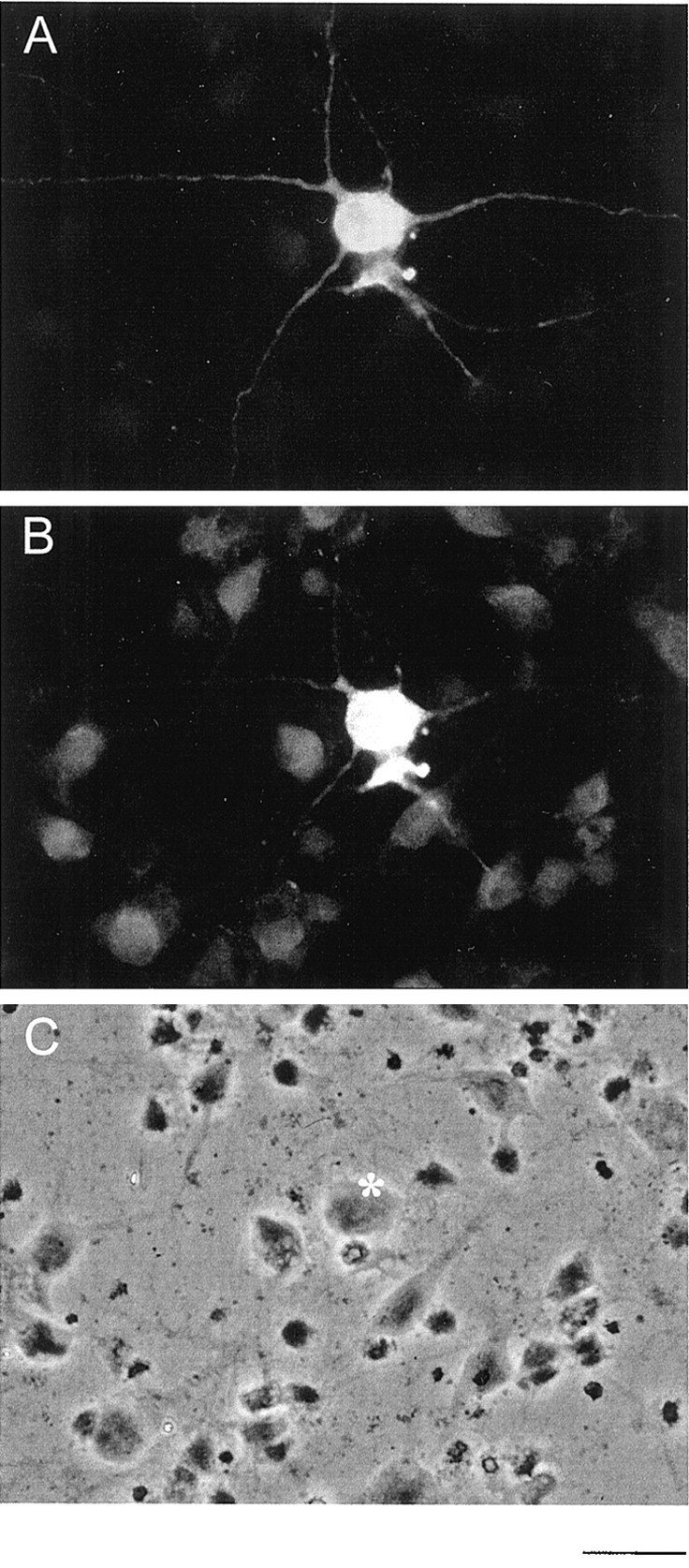
Immunofluorescence microscopic analysis of serotonergic neurons in raphe primary culture. Raphe neurons cultivated for 10 d in vitro were fixed and incubated using a mixture of chicken anti-serotonin antiserum and rabbit anti-VMAT2 antiserum. Of a couple of neurons shown in C, only one neuron labeled by an asterisk was positively stained for serotonin (A) or VMAT2 (B). Scale bar, 10 μm.