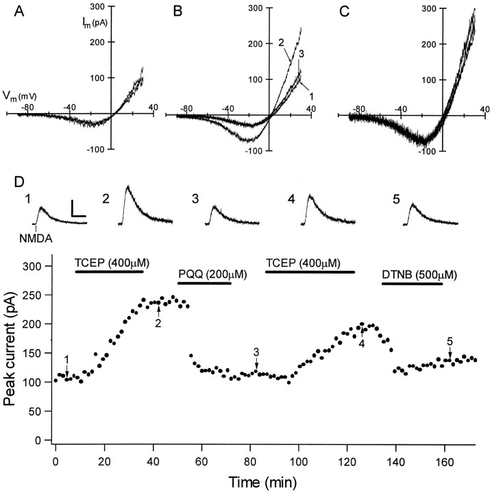
Fig. 4.
NMDA-evoked whole-cell currents are modulated by redox reagents at concentrations that modulated epileptiform activity. A,I–V curves for NMDA-evoked currents were obtained by subtracting the responses to command voltage ramps from −90 to +30 mV before and during brief NMDA superfusion. Subtracted current responses revealed characteristic voltage-dependent currents that reversed between 0 and +10 mV, and the superimposed subtracted ramps for three consecutive NMDA applications show the reproducibility of responses obtained in this manner.B, Superimposed I–V curves show that baseline NMDA-evoked currents (trace 1) were significantly potentiated by exposure to 500 μm TCEP (trace 2) and subsequently diminished by 200 μm PQQ (trace 3). Eachtrace is an average of three subtracted ramp responses.C, Ramp currents from B scaled to the largest current at −20 mV showed that TCEP and PQQ exerted modulation with no effect on the voltage dependence or reversal potential of the NMDA-evoked currents. D, Persistence of the effects of redox reagents on currents evoked by focal NMDA application are shown. The graph shows the peak amplitude of individual current responses to focal NMDA application (holding potential = +30 mV) as a function of time for a CA1 pyramidal neuron that was recorded for 3 hr. (Horizontalbars show the time of superfusion of each redox agent.) The initial application of 400 μm TCEP caused a gradual increase in the peak NMDA-evoked current that remained unchanged during TCEP washout but very rapidly returned to baseline after subsequent exposure to 200 μm PQQ. Responses were potentiated again by a second TCEP application, and this effect was reversed by exposure to 500 μm DTNB, mimicking the effect of PQQ. Individualtraces are shown at the top for thepoints labeled in the graph. Calibration: vertical, 100 pA; horizontal, 10 sec.

