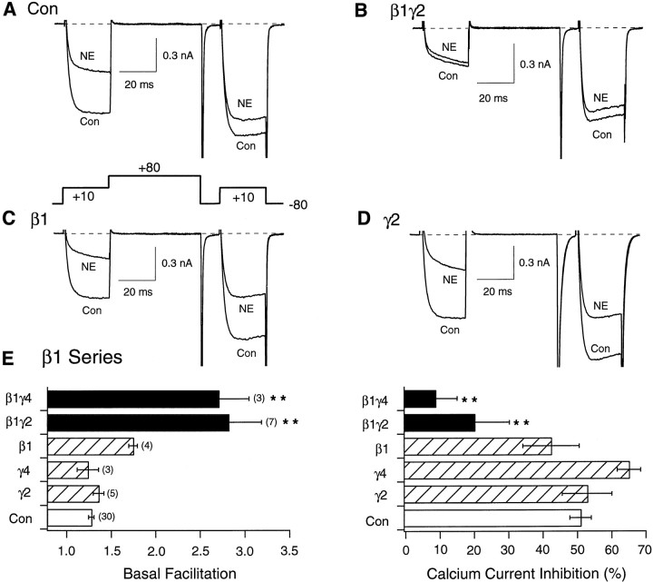
Fig. 1.
Facilitation and NE-mediated inhibition of Ca2+ currents in SCG neurons expressing β1 or Gγ alone or combined. Superimposed Ca2+ current traces evoke with the “double-pulse” voltage protocol (bottom of A) in the absence (bottom traces) and presence (top traces) of 10 μm NE for control (A), Gβ1γ2- (B), Gβ1- (C), and Gγ2-expressing (D) neurons. Currents were evoked every 10 sec. E, Summary graphs of mean ± SEM basal facilitation and Ca2+ current inhibition for neurons expressing Gβ1 alone or combined with Gγ2 and Gγ4 subunits. Final concentration of cDNA injected was 10 ng/μl per subunit. Facilitation was calculated as the ratio of Ca2+ current amplitude determined from the test pulse (+10 mV) occurring after (postpulse) and before (prepulse) the +80 mV conditioning pulse. Ca2+ current inhibition was measured isochronally 10 msec after initiation of the test pulse (+10 mV) in the absence or presence of 10 μm NE. **p < 0.01 versus control. Numbersin parentheses indicate the number of experiments.

