
Fig. 1.
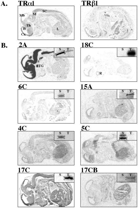
Distribution of putative thyroid hormone-responsive genes in the GD16 embryo. Images are derived from film autoradiograms after in situ hybridization to determine whether putative thyroid hormone-responsive mRNAs identified by differential display RT-PCR were expressed in areas known to contain TR mRNAs (Experiment I). RNA probes were applied to sagittal sections of GD16 rat embryos. Sense controls were applied to adjacent sections and produced negligible hybridization signal (data not shown), with the exception of fragment 17C, which we did not examine further.A, Distribution of TRα1 and TRβ1 mRNA.B, Distribution of putative thyroid hormone-responsive mRNAs as noted above panel. Film autoradiograms of gene fragments identified by differential display are shown in insets, illustrating direction of thyroid hormone effects. See Table 1 for RNAimage primers used to generate each fragment. Cx, Cortex; H, hippocampus; L, liver;M, medulla; Mb, midbrain;R, retina; S, saline-injected;SC, spinal cord; T, T4-injected; TG, trigeminal ganglion;Th, thalamus. Scale bar, 0.5 cm.