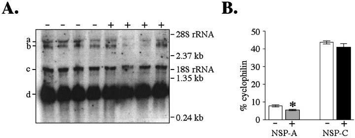
Fig. 4.
Effect of thyroid hormone manipulation on the expression of NSP variants using quantitative Northern analysis (Experiment II). A, Phosphor-image of Northern blot in which 20 μg total RNA was hybridized with 32P-labeled NSP cDNA. 32P-labeled cyclophilin cDNA was hybridized simultaneously to control for loading variations. RNA was isolated from GD16 fetuses derived from hypothyroid dams receiving either no injection (−) or a single T4 injection at 9 P.M. on GD14 (+). Each sample corresponds to RNA pooled from four or five animals. Positions of the molecular weight standards and the 18S and 28S ribosomal RNAs are indicated. B, Quantification of band density shown in A using ImageQuant software. NSP-A mRNA was decreased after acute maternal T4 exposure. Bars represent mean band density ± SEM (converted to % total cyclophilin). a Transcript of 3.5 kb hybridized to NSP probe, corresponding to NSP-A. b Transcript of 3.4 kb hybridized to cyclophilin probe. c Transcript of 1.5 kb hybridized to NSP probe, corresponding to NSP-C.d Transcript of 0.9 kb hybridized to cyclophilin probe. *Significantly different from hypothyroid dams receiving no injection (p < 0.05).

