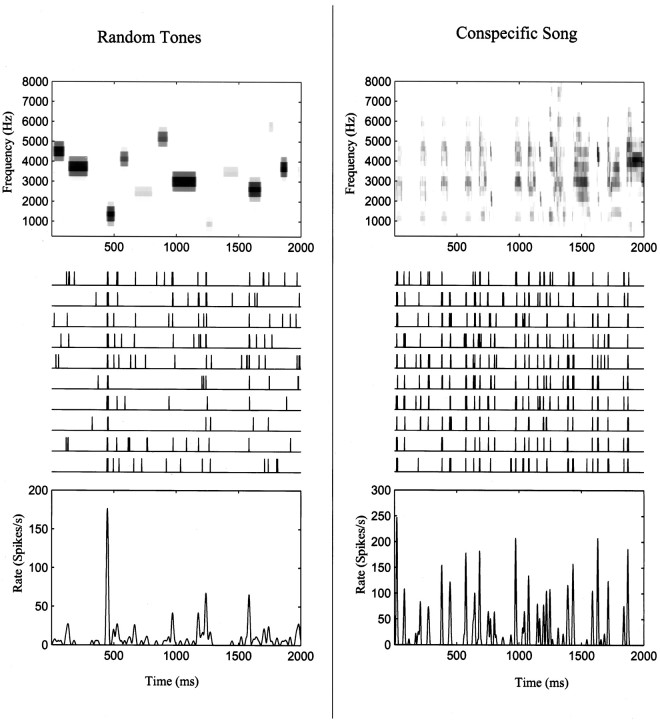
Fig. 3.
Samples of stimuli (top panels), neuronal responses (middle panels), and mean spike rate (bottom panels) for the two stimulus ensembles: a song-like sequence of random tone pips (left column) and a zebra finch song (right column). The top panels show the spectrographic representation of the stimuli using the frequency decomposition described in Materials and Methods. Themiddle panels show 10 spike train recordings obtained in response to 10 presentations of the stimuli for a neuronal site in area L2b of one of the birds used in the experiment. The bottom panel shows the estimated spike rate obtained by smoothing the PSTH with a 6 msec time window.

