
Fig. 6.
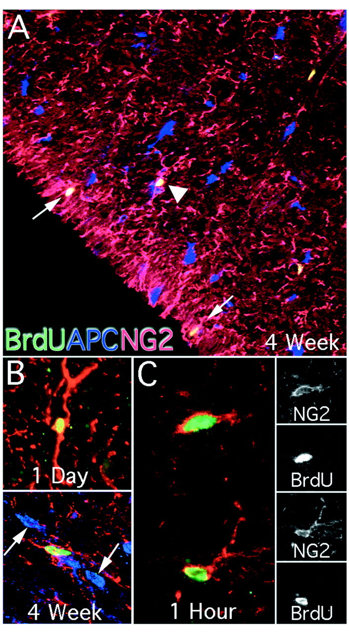
NG2 colocalization with BrdU in the adult spinal cord. NG2 immunoreactivity was used to classify BrdU-positive cells as glial progenitor cells. NG2 immunoreactivity was found throughout the spinal cord and labeled cells exhibited unipolar, bipolar, and multipolar cell morphologies in both gray and white matter. NG2 colocalized with BrdU more commonly than any other immunohistochemical marker. Throughout the spinal cord BrdU/NG2 colocalized cells were found that did not colabel with the glial marker APC (A, arrowhead, 100×). Many BrdU/NG2-colabeled cells were located near the pial layer with processes extending into this region (A, arrows). These cells were not found near the central canal. The predominant morphology was that of a bipolar cell (B, top, 400×). At 4 weeks, a small population had complex, multipolar processes (B, bottom, 400×). These cells did not colocalize with the mature glial marker APC. At 1 hr after a single pulse of BrdU, most BrdU-labeled cells colocalized with NG2 (C, 630×). Single confocal sections of each marker are presented for both NG2-immunoreactive cells.