
Fig. 7.
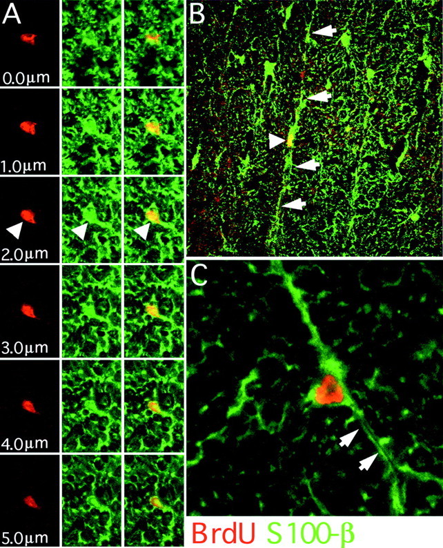
BrdU/S100β colocalization in the adult spinal cord. Confocal microscopy was used to determine the incidence of BrdU/S100β colocalization. A confocal z-series allows examination of nuclei through their entire z-axis in 1 μm steps (A, 400×). In this series a BrdU-positive nucleus (red only) is associated with S100β immunoreactivity (green only). These markers consistently colocalize (red and greenmerge) throughout the series (arrowheads). S100β astrocytes (arrowhead) often exhibited a radial morphology with long central to lateral processes (B,arrows, 200×). Occasionally BrdU-positive astrocytes had processes that contained a lumen associated with microvascular elements (C, arrows, 800×).