
Fig. 8.
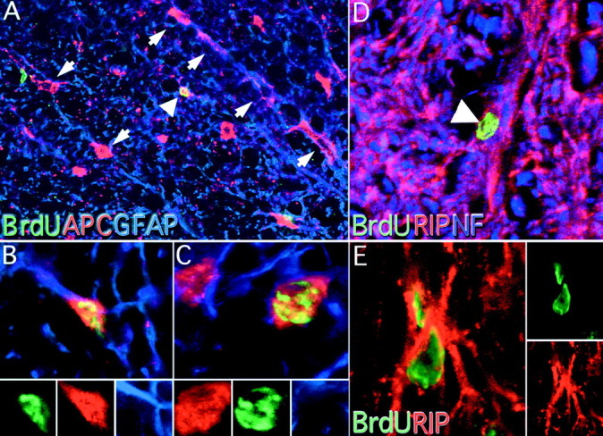
APC, GFAP, and RIP colocalization with BrdU in the adult spinal cord. APC and GFAP immunoreactivity were used to classify BrdU-positive cells into immature oligodendrocytes or astrocytes. APC immunoreactivity was found throughout the spinal cord, especially in the white matter where astrocytes and oligodendrocytes were found in radially oriented chains (A, arrows, 200×). Astrocytes were characterized by a small somal size and colocalization with GFAP (A, arrowhead;B, 800×). Oligodendrocytes were also detected with this method. Oligodendrocytes contained large, rounded cell bodies that did not colocalize with GFAP (C, 8000×). Separate color channels are presented at the bottom of B andC. Mature oligodendrocytes were identified by colocalization of BrdU and RIP immunoreactivity (D, arrowhead, 400×). Confocal microscopy was used to determine if RIP-positive cell bodies contained BrdU-positive nuclei (E, 800×). Separate confocal channels are presented to the right.