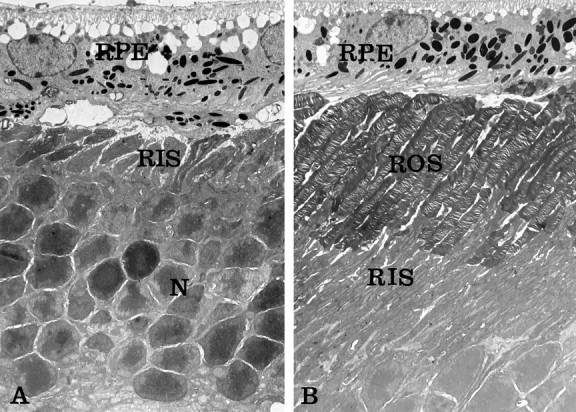
Fig. 4.

Electron micrographs of 3-month-old mice.A, Homozygous rds retina expressing Bcl-2 (rds:bcl). Note the absence of outer segments and the abnormal position of photoreceptor rod inner segments (RIS) against the retinal pigment epithelium (RPE). The effect of Bcl-2 on photoreceptor survival is evident by the presence of a significant number of photoreceptor nuclei (N). B, Homozygousrds retina expressing both normal rds/peripherin and Bcl-2 transgenes (rds:113+bcl). Note the normal morphology of intact rod outer segments (ROS) adjacent to the RPE. The expression of Bcl-2 did not adversely affect photoreceptor organization. Magnification: A, 3200×; B, 2900×.
