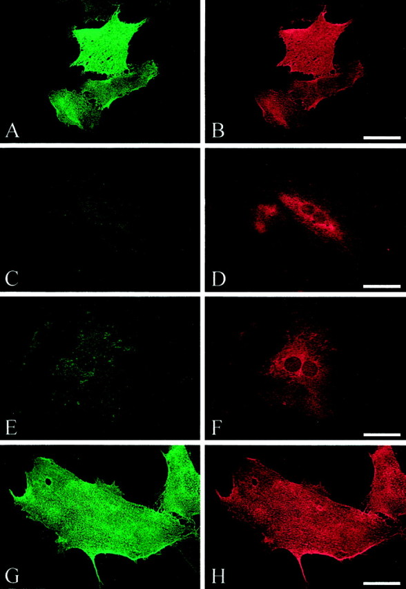
Fig. 3.

Surface (A, C,E, G) and permeabilized (B, D, F,H) staining of L1. Double immunofluorescence was performed on rat cerebellar astrocytes infected with dvL1 (16.5 hr after infection): wild type, A, B; R184Q,C, D; D598N, E,F; and S1194L, G, H. In most cells expressing R184Q and D598N mutant L1, L1 protein failed to reach the cell surface. Scale bar, 50 μm.
