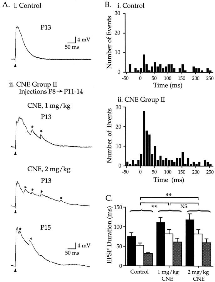
Fig. 3.
CNE group II: nicotine treatment during postnatal week 2 alters EPSPs. Ai, Representative EPSP from a control P13 neuron; Vm −73 mV.Aii, Nicotine injections (1 or 2 mg/kg) from P8 through P11–14 produced EPSPs that were larger in duration and magnitude and had multiple peaks on their descending phase (asterisks). Top trace,Vm −66 mV; middle trace,Vm −66 mV; bottom trace,Vm −77 mV. B, The time of occurrence of miniature fluctuations is plotted for 50 msec before and 260 msec after the stimulus (10 msec bins; comparisons using Fisher's PLSD). Bi, In 22 control neurons (P9–15), the number of events occurring before or 60 msec after the stimulus did not differ (p > 0.10). Bii, For 22 CNE group II neurons (1 and 2 mg/kg doses combined), the number of events within 60 msec after the stimulus was greater than during either the prestimulus period or the same 60 msec poststimulus period in control neurons (p > 0.10). C, EPSP durations recorded from CNE group II neurons (measured at , ½, and peak; histogram shading as in Fig. 2) were longer than age-matched controls (Table 1; p < 0.01). The increase in duration was similar for the two nicotine doses (p > 0.10).

