
Fig. 1.
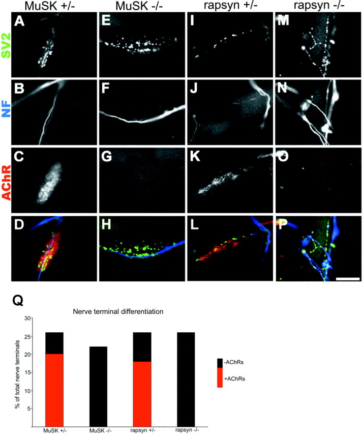
Nerve–muscle cocultures. A–P, Fluorescent photomicrographs of nerve–muscle cocultures showing terminal differentiation of neurites as evidenced by the presence of SV2 staining (A, E, I, M) in neurofilament-poor (B, F, J, N) areas of the nerve terminals. Twenty-six percent of nerve–muscle contacts in control cocultures (A, B, I, J) showed signs of differentiation in that the neurites formed a small array of neurofilament-poor, SV2-rich branches. Approximately 73% of these differentiated contacts were found overlying high-density AChRs labeling with TRITC-BTX (C, K), whereas the remaining 27% of the contacts were on AChR-poor membranes. A similar degree of presynaptic differentiation as evidenced by neurofilament-poor, SV2-rich branches was seen in cocultures with MuSK−/− (E, F) and rapsyn−/− (M, N) myotubes. All of these differentiated nerve–muscle contacts in cocultures with mutant muscle fibers occurred on AChR-poor membranes (G, O). D, H, L, P, Superimposition of presynaptic and postsynaptic staining for SV2 (pseudocolored green), neurofilament (pseudocoloredblue), and AchRs (peudocolored red) for control and mutant cocultures. Scale bar, 5 μm. Q, Column graph showing that the level of nerve terminal differentiation is similar in the control and mutant muscle fiber cocultures.