
Fig. 2.
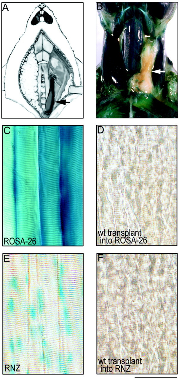
Transplantation method. A, Schematic of transplantation method showing a sternomastoid muscle from a P0 mouse (arrow) transplanted into the neck of an adult mouse whose own sternomastoid muscle has been removed.B–D, Wild-type muscle grafted into a ROSA-26 host 4 weeks after transplantation. B, Dissected neck of a ROSA mouse whose own left sternomastoid muscle has been replaced with a wild-type muscle graft (arrow) 1 month after transplantation. C, Nearly all cell types in the ROSA mice, including skeletal muscle fibers, express cytoplasmic β-galactosidase and turn blue with X-gal staining.D, In contrast, wild-type (wt) muscle fibers grafted into ROSA-26 hosts remain completely clear. E, F, Wild-type muscle grafted into an RNZ host. E, Muscle fibers in the RNZ mice express nuclear localized β-galactosidase, and thus the myonuclei in these mice turnblue with X-gal staining. F, In contrast, wild-type muscle fibers grafted into the RNZ mice show complete absence of blue myonuclei. These results indicate that there has been no fusion of host precursor cells with muscle fibers in the graft. Scale bar (B–E), 100 μm.