
Fig. 4.
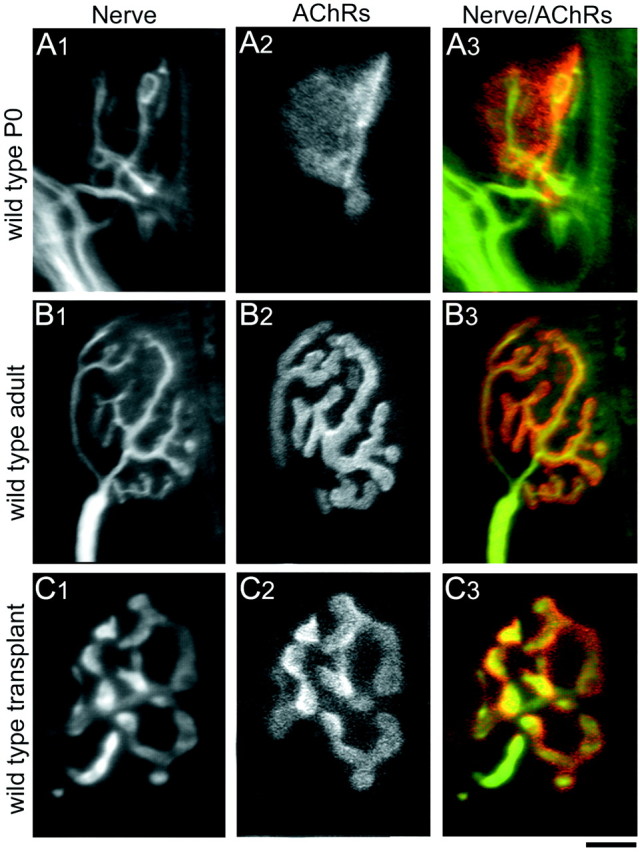
Comparison of NMJs from wild-type P0, adult, and transplanted muscle fibers. A, At P0, nerve terminals (A1, immunolabeled with antibodies against neurofilament and synaptophysin) have small bouton-like endings overlying a plaque-like area of high AChR density (A2, labeled with TRITC-BTX; A3, overlay of nerve terminal pattern on AChR pattern). B, By 4–6 weeks postnatally in mice, each muscle fiber receives innervation from a single myelinated axon whose arbors show extensive branching (B1). The postsynaptic AChR cluster underlying the presynaptic nerve terminals also shows breaking apart of the original plaque to form a high, branched structure (B2). The presynaptic nerve terminals and the postsynaptic AChR clusters correspond exactly in their branching pattern (B3). C, Four weeks to 10 months after transplantation, wild-type muscle fibers are also innervated by a single axon. The overall size of the NMJs in transplanted muscle fibers is smaller compared with normal adult NMJs, probably because of the fact that the neonatal graft was placed in a full sized adult host, and thus the muscle fibers themselves do not lengthen and are consequently smaller than their age-matched nontransplanted counterparts. The presynaptic (C1) and postsynaptic (C2) components of the NMJs in wild-type transplanted muscle fibers exactly oppose each other (C3) and show the branching morphology similar to that seen in normal nontransplanted NMJs. The difference in presynaptic arbor width between B1 and C1reflects variability in antibody staining for synaptophysin and does not reflect differences in presynaptic structure between wild-type adult and wild-type transplants. Scale bar: A, C, 5 μm; B, 12.5 μm.