
Fig. 7.
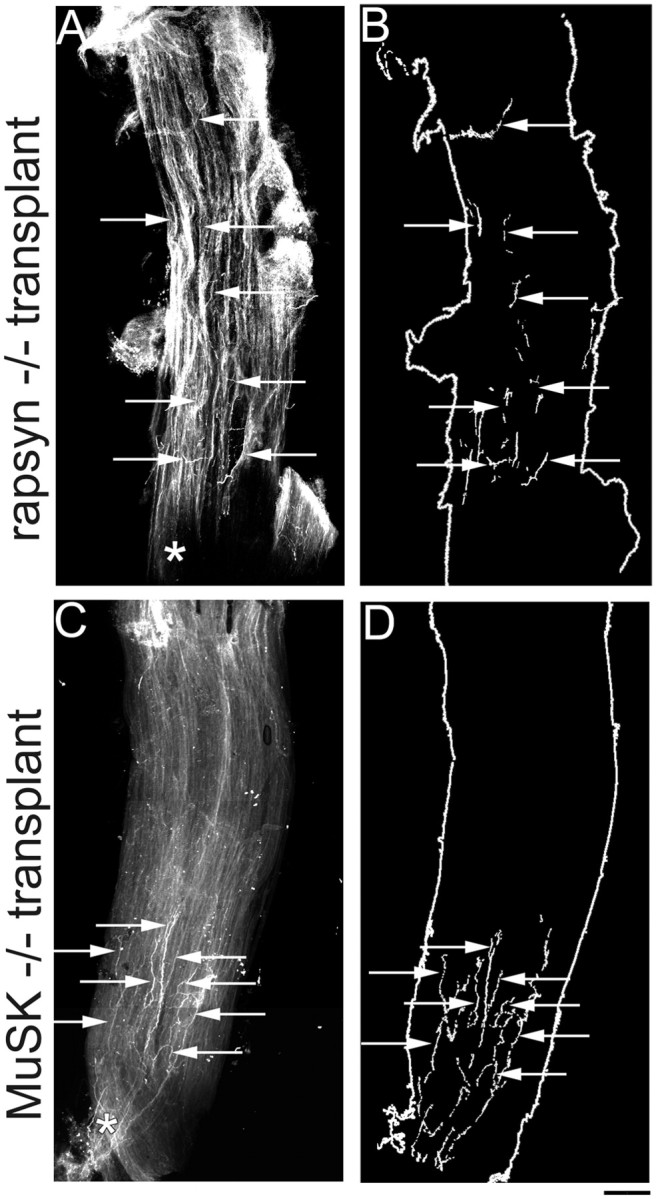
Distribution of host axons in rapsyn−/− and MuSK−/− grafts. A, Low-power photomicrograph showing an example of the innervation pattern of a rapsyn−/− muscle after transplantation into a wild-type host (11 weeks after transplantation). Normal host axons innervating rapsyn−/− muscles terminate far away from the entry point of the intramuscular nerve (asterisk). B, Schematic of rapsyn−/− graft in A, showing that nerve terminal endings (arrows) can be found throughout the length of the graft. (Note: the fragmented appearance of the nerve terminals is attributable to the fact that innervating axons weave between the muscle fibers of the graft and thus disappear from the plane of focus.)C, Low-power photomicrograph showing an example of a MuSK−/− muscle after transplantation into a wild-type host (7 months after transplantation). In contrast to the diffuse innervation pattern of muscles in MuSK−/− mutants at P0, after transplantation, wild-type axons innervating MuSK−/− muscles terminate closely to the entry point of the intramuscular nerve (asterisk).D, Schematic of the MuSK−/− graft shown inC, showing that nerve terminal endings (arrows) are found in a tight cluster near the middle of the graft along its length. Scale bar, 200 μm.