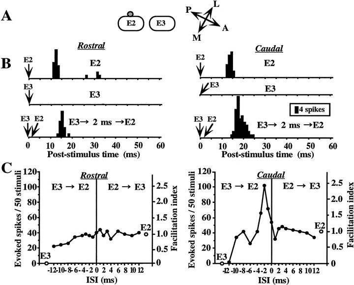
Fig. 4.
Example of a layer II/III cell showing direction-selective facilitation. A, The cell was located on the side of the E2 barrel column. B, PSTHs of responses to deflections of E2 and E3. PSTHs in the left column, Whiskers were deflected in the rostral direction.PSTHs in the right column, Whiskers were deflected in the caudal direction. Top and middle, Responses to single whisker stimulation of E2 and E3, respectively.Bottom, E3 stimulation preceded E2 stimulation by 2 msec. Facilitation (243%) was observed for the response to caudal deflection. Other notations are as in Figure 2. Note that both facilitatory and suppressive interactions were direction selective, whereas responses to single whisker stimulation were almost the same for the two directions (open circles ingraphs, and B, middle andbottom).

