Fig. 8.

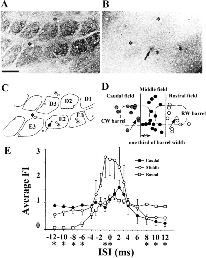
Relationship between response interaction and cell location in relation to the barrel structure in the superficial layers.A, B, Tangential sections of layer IV showing barrels (A) and layer II/III (B) of the PMBSF of a recorded animal. The section was stained for cytochrome oxidase. Arrow and asterisks indicate a dye mark of Pontamine Sky Blue produced at the recording site and blood vessels used as references for the reconstruction, respectively. Scale bar, 500 μm. C, Recording site and barrel arrangement reconstructed by superimposing the image of B on that ofA. D, Tangential reconstruction of the distribution of cells. The barrels in layer IV are superimposed in dashed lines. Cells were divided into three groups based on the location in relation to two barrel columns; “caudal”, “middle”, and “rostral” field groups. Middle field group consisted of cells located adjacent to one-third of two barrels and the septal area.E, Averaged facilitation indices of each group for various ISIs (mean ± SE). Asterisks below the ISIs mean that there are statistically significant differences among the groups (one-way ANOVA, p < 0.05).
