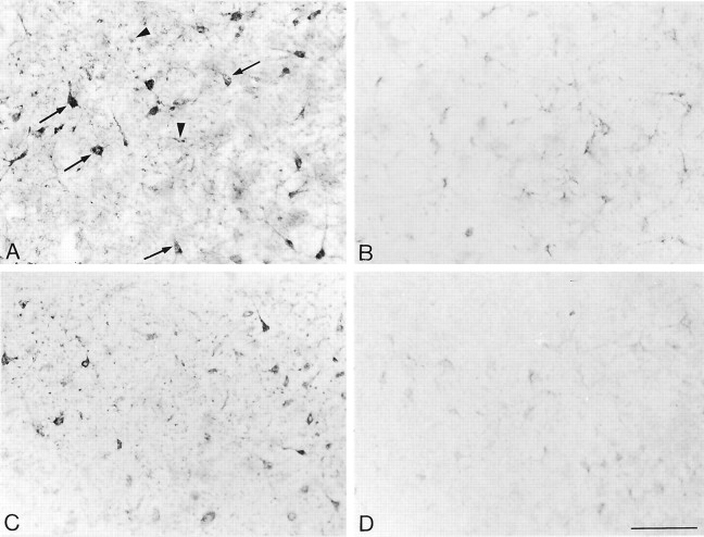
Fig. 1.
Light microscopic distribution of SST2A receptor immunolabeling in claustrum slices incubated for 40 min at 37°C in Ringer's buffer (A), in Ca2+-free buffer (B), in Ca2+-free buffer containing 100 nm[d-Trp8]-SRIF (C), and in Ca2+-free buffer containing 100 nm[d-Trp8]-SRIF plus 10 μm PAO (D). A, After incubation in Ringer's, intense immunolabeling is observed over nerve cell bodies and their proximal dendrites (arrows). Punctate immunostaining typical of cross-sectioned immunoreactive dendrites is also evident (arrowheads).B, After incubation in Ca2+-free buffer, the intensity of SST2A immunolabeling is reduced dramatically in both cell bodies and surrounding neuropil.C, Addition of 100 nm[d-Trp8]-SRIF to the Ca2+-free incubation medium almost totally reestablishes the level of SST2A immunoreactivity to that seen in Ringer's controls. D, [d-Trp8]-SRIF-induced recovery of SST2A immunoreactivity is totally prevented by addition of the endocytosis inhibitor PAO to the incubation medium. Scale bar, 100 μm.

