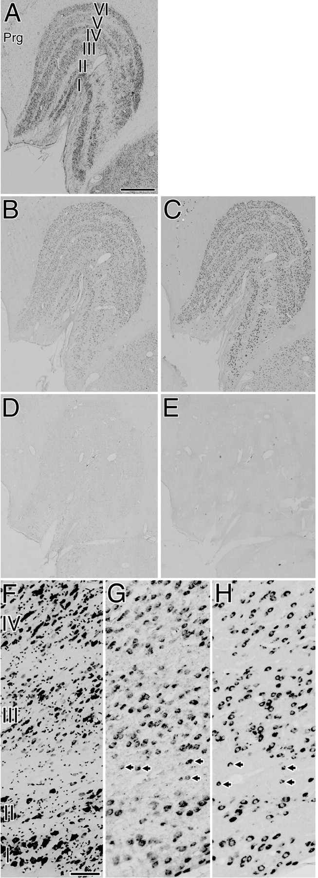
Fig. 1.

A–E, Five adjacent sections of the normal LGN. Expression of both GAP-43 and SCG10 mRNAs was observed in the neurons of all layers. The expression patterns of both GAP-43 and SCG10 mRNAs were similar among all animals examined and throughout the rostral to caudal levels of the coronal sections.A, Nissl-stained section. B, Localization of GAP-43 mRNA. C, Localization of SCG10 mRNA.D, Control section hybridized with a sense probe for GAP-43. E, Control section hybridized with a sense probe for SCG10. Prg, Perigeniculate nucleus. Scale bar, 1 mm.F–H, Higher-magnification photomicrographs. F, Nissl-stained sections. G, Localization of GAP-43 mRNA.H, Localization of SCG10 mRNA. Arrowsindicate GAP-43 or SCG10 mRNA-positive neurons in the interlaminar regions. Many GAP-43 or SCG10 mRNA-positive neurons existed in magnocellular layers (layers I and II), parvocellular layers (layers III–VI), and interlaminar regions. Scale bar, 100 μm.
