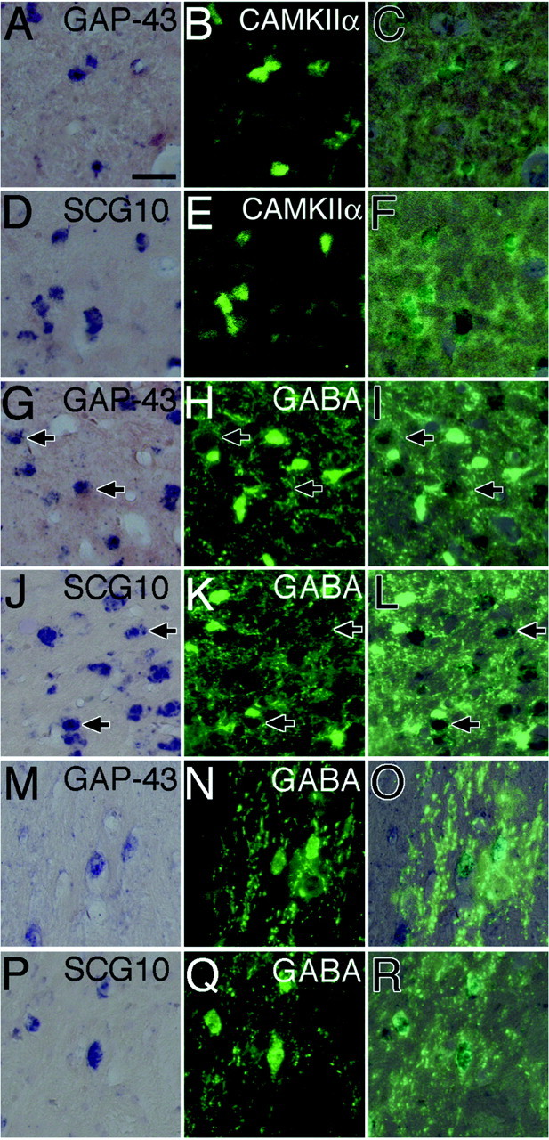
Fig. 3.

A–F, Expression of GAP-43 and SCG10 mRNAs in CAMKII-α-immunoreactive neurons (koniocellular neurons) in the interlaminar region between layers I and II of the LGN. A, Localization of GAP-43 mRNA.B, Localization of CAMKII-α in the same section asA. C, The image of B was superimposed on the image of A. CAMKII-α-immunoreactive neurons (green) expressed GAP-43 mRNA (dark blue).D, Localization of SCG10 mRNA. E, Localization of CAMKII-α in the same section as D.F, The image of E was superimposed on the image of D. CAMKII-α-immunoreactive neurons (green) expressed SCG10 mRNA (dark blue). G–L, Absence of GAP-43 and SCG10 mRNAs in GABA-immunoreactive neurons in layer II of the LGN.Arrows indicate GAP-43 or SCG10 mRNA-positive neurons that were outlined by GABA-immunoreactive terminals. G, Localization of GAP-43 mRNA. H, Localization of GABA in the same section as G. I, The image ofH was superimposed on the image of G. GABA-immunoreactive neurons (green) did not express GAP-43 mRNA (dark blue). J, Localization of SCG10 mRNA. K, Localization of GABA in the same section as J. L, The image ofK was superimposed on the image of J. GABA-immunoreactive neurons (green) did not express SCG10 mRNA (dark blue).M–R, Expression of GAP-43 and SCG10 mRNAs in GABA-immunoreactive neurons in the perigeniculate nucleus.M, Localization of GAP-43 mRNA. N, Localization of GABA in the same section as M.O, The image of N was superimposed on the image of M. GABA-immunoreactive neurons (green) expressed GAP-43 mRNA (dark blue). P, Localization of SCG10 mRNA.Q, Localization of GABA in the same section asP. R, The image of Q was superimposed on the image of P. GABA-immunoreactive neurons (green) expressed SCG10 mRNA (dark blue). Scale bar, 50 μm.
