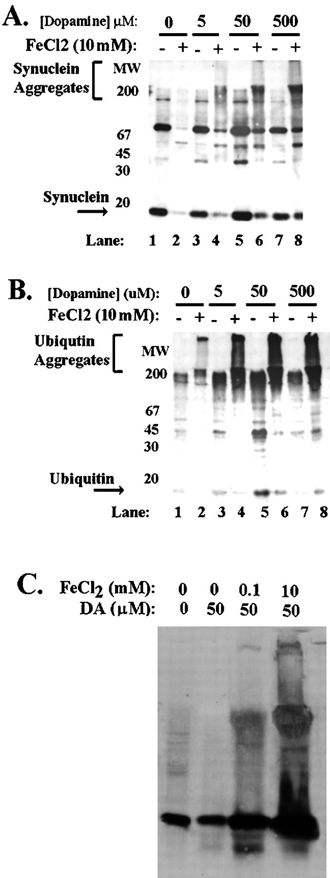
Fig. 2.

Free radicals potentiate induction of α-synuclein aggregation by iron. A, B, Aggregation of α-synuclein in BE-M17 cells expressing A30P α-synuclein after treatment with 0, 5, 50, and 500 μmdopamine plus or minus 10 mm FeCl2 for 48 hr. The immunoblot was first probed with monoclonal anti-synuclein antibody (A) and then reprobed with anti-ubiquitin antibody (B). C, Immunoblots of lysates from primary cortical neurons after treatment with 0, 0.1, or 10 mm FeCl2 and 50 μm dopamine (DA) for 60 hr.
