Fig. 6.

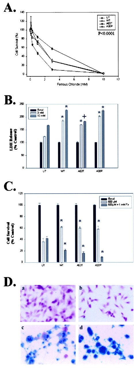
α-Synuclein increases iron-dependent toxicity.A, B, BE-M17 cells overexpressing wild-type, A53T, or A30P α-synuclein were treated with varying doses of FeCl2 for 48 hr, and the viability was determined using the MTT assay (A) or the LDH assay (B). C, BE-M17 cells overexpressing wild-type, A53T, or A30P α-synuclein were treated with varying doses of H2O2 for 48 hr, and the viability was determined using the MTT assay. *p < 0.001, +p < 0.01 by ANOVA analysis.D, Iron stain of BE-M17 cells. a, Untransfected cells, basal conditions; b, A53T α-synuclein-expressing cells, basal conditions; c, untransfected cells, FeCl2–H2O2; d, A53T α-synuclein-expressing cells, FeCl2–H2O2. Under basal conditions, none of the cells showed staining for iron, but after treatment with 10 mm FeCl2 and 100 μm H2O2 for 48 hr, the A53T cells showed much more iron reactivity (blue, iron stain;pink, nuclear–cytoplasmic counterstain).
