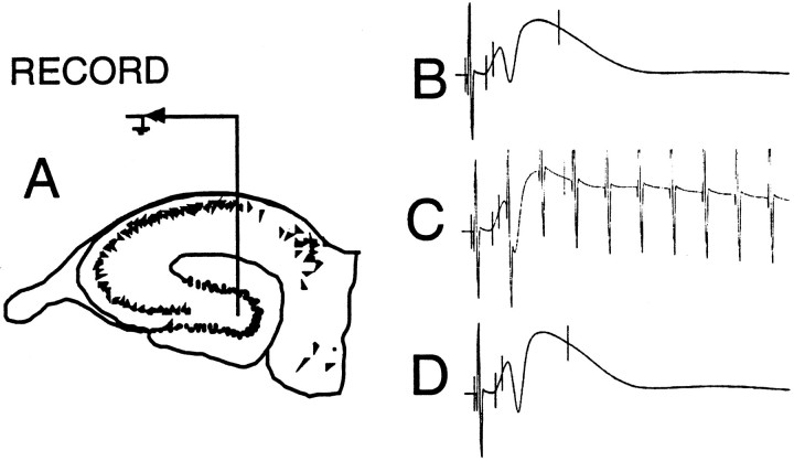
Fig. 1.
A, Schematic illustration of the placement of the chronically implanted infusion cannula/recording electrode in the hilus of the fascia dentata. The stimulating electrode was implanted in the angular bundle to optimize the perforant path–granule cell evoked field potential. B, Example of the extracellular hippocampal evoked response elicited from perforant path stimulation before high-frequency stimulation. C, Example of the response during the high-frequency burst used to induce LTP. D, Example of the response on the day after LTP induction.

