
Fig. 1.
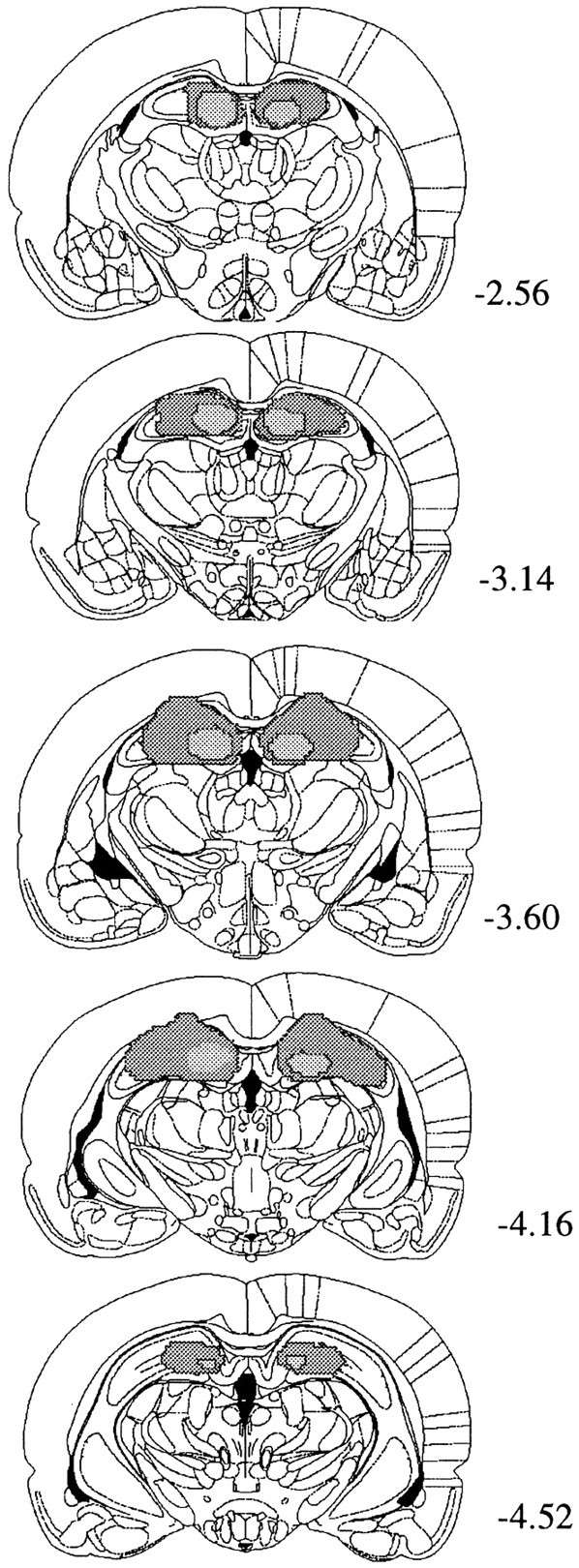
Line drawings of coronal sections from the brains of subjects with the maximum and minimum damage resulting from lesions in the hippocampal group. Starting from the top, sections are taken from the following points in the anteroposterior plane (in millimeters relative to bregma): −2.56, −3.14, −3.60, −4.16, and −4.52. Drawings are from Paxinos and Watson (1998).