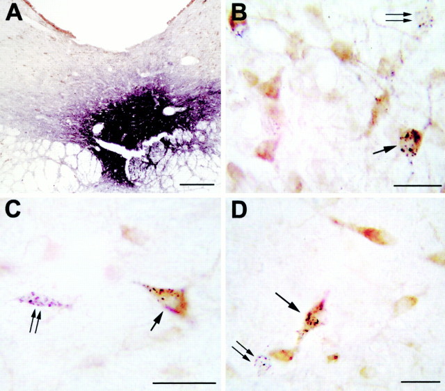
Fig. 7.
A, Photomicrograph illustrating a small CTb injection site limited to the ventral part of the DRN. Scale bar, 300 μm. B–D, Photomicrographs showing GAD (light brown) and CTb (black granules) double-labeled neurons (arrow), singly labeled GAD immunoreactive neurons, and singly CTb-labeled neurons (double arrow) in the lateral preoptic area (B), the lateral hypothalamic area (C), and the ventral periaqueductal gray (D) after a CTb injection in the DRN. Scale bars, 25 μm. Note that the singly CTb-labeled cells display no brown coloration, indicating the absence of cross-reactivity between our two immunohistochemical reactions.

