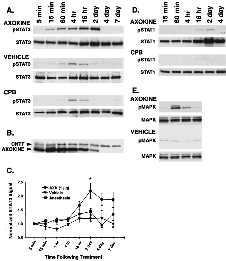
Fig. 1.
Effects of Axokine on phosphorylated and total STAT3, STAT1, and MAPK in homogenized rat retinas. A, Retinas were harvested at various times indicated (5 min to 7 d) after a single intravitreal injection of Axokine. Homogenized retinas were Western blotted (50 μg/lane) to detect total STAT3 and Y705-phosphorylated STAT3 (pSTAT3). The band size for both STAT3 and pSTAT3 is the expected 92 kDa. Axokine produced robust pSTAT3 signal beginning at 15 min and disappearing by 4 d. Weaker and more transient pSTAT3 signals were seen in the vehicle and anesthesia (CPB) control groups. B, Membranes from Axokine-treated STAT3/pSTAT3 immunoblots were stripped and reblotted using an antibody that recognizes both CNTF and Axokine (23 and 21 kDa, respectively). Axokine was detected in the retina for up to 2 d after injection. C, Graph summarizing the time-dependent effects of total STAT3 for each treatment group. STAT3 signal was quantified at each time point using conventional densitometric analysis and normalized to the STAT3 signal at 5 min. Analysis of immunoblots from time course studies culled from three separate groups of animals shows a significant increase in normalized STAT3 signal in the Axokine-treated group (p < 0.05; repeated ANOVA, Dunnett's post hocttest). Error bars are in terms of SE. D, Immunoblots for total STAT1 and Y701-STAT1 (pSTAT1) were performed on Axokine-treated and anesthesia control retinas. Axokine weakly activated pSTAT1 at the later time points but dramatically increased total STAT1 levels by 16 hr. E, Western blots for total MAPK and pMAPK for Axokine-treated retinas showed robust transient activation of MAPK at 60 min.

