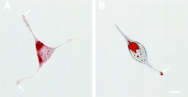
Fig. 5.
Confocal microscopy of AtT-20 (A) and AtT-20/α1-PDX cells (B) infected with VV encoding pro-NT-3. Cells were infected for 1 hr and postincubated in the absence of virus for another 8 hr. The cultures were fixed and treated with antibodies against NT-3 (Chemicon), followed by CY3-conjugated goat anti-rabbit IgG. Scale bar, 10 μm. Photomicrographs were obtained by overlaying confocal fluorescence images over transmitted light images. Scale bar, 10 μm.

