
Fig. 3.
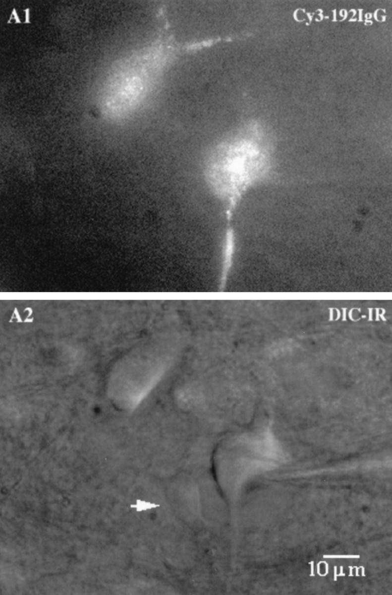
Visualization of septohippocampal cholinergic neurons in live rat brain slices using Cy3–192IgG, a selective marker of p75-receptor-expressing neurons in the MSDB. A,Cy3–192IgG-labeled neurons show a granular fluorescent labeling as seen in a 300-μm-thick slice preparation. B, The same neurons visualized using differential interference contrast, infrared videomicroscopy. Note an unlabeled neuron (arrow) and the healthy appearance of the labeled cells.