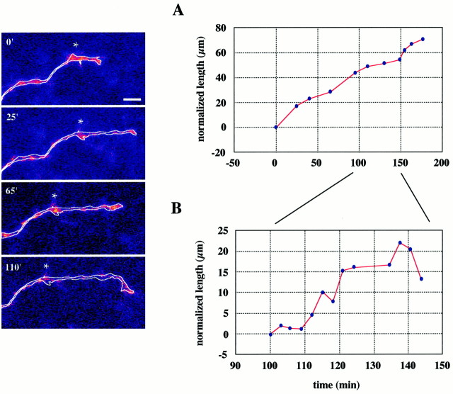
Fig. 3.
E16 thalamic fiber growing through the IC.Side panels are extended-focus images of the distal segment of the axon and its growth cone, collected at various times throughout a 3 hr imaging session. The axon has beenoutlined for clarity. The times at which the stacks were collected are indicated at the top left of each image, and the star indicates a stable point along the axon for clearer illustration of axon extension. The top graphshows that it advanced at a relatively constant growth rate. Thebottom graph, at an expanded time scale, provides a better view of the momentary pauses and retractions. Scale bar, 10 μm.

