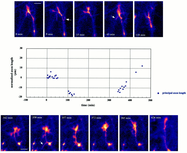
Fig. 4.
Thalamic axon with its growth cone located in the ventral intermediate zone (VIZ) of an E17 forebrain slice.Panels at the top andbottom are single confocal frames obtained at the times indicated. These images are framed to illustrate growth cone morphology and do not necessarily reflect the absolute position of the growth cone with respect to the slice. The graph illustrates the behavior of this axon for the 8 hr of the imaging session. Measurements were made from a stable inflection point further down along the axon shaft and were normalized to the beginning of the sequence.Dots indicate the principal axon length, i.e., the distance from the measuring point to the leading edge of the body of the growth cone (excluding filopodia and side branches, examples of which are indicated by the arrows of the leading segment of the axon). Periods lacking data points reflect either loss of the focal plane or deliberate changes in the area that was imaged. Although the out-of-focus images were not sharp enough to allow for accurate measurements, they confirmed that the growth cone was still in the field of view and had not manifested massive extensions or retractions. Note that the net forward growth rate is at its highest at the end of the 8 hr imaging session, indicating the continuing good health of the slice. Scale bar, 10 μm.

